Home
/ Pelvic Anatomy Xray - Abdomen And Pelvis Radiographs And Images Xrays And _ What is the collateral circulation after hypogastric artery ligation?
Pelvic Anatomy Xray - Abdomen And Pelvis Radiographs And Images Xrays And _ What is the collateral circulation after hypogastric artery ligation?
Pelvic Anatomy Xray - Abdomen And Pelvis Radiographs And Images Xrays And _ What is the collateral circulation after hypogastric artery ligation?. Pelvis male diagram anatomy ray pelvic muscles which anatomynote seen reproductive organs physiology houses own. It is subdivided into the greater pelvis and lesser pelvis. Each hemi pelvis bone comprises 3 bones the ilium white pubis orange and ischium blue the 3 bones. ●to review pelvic sidewall anatomy including retroperitoneal spaces. The geometry of bony pelvis differs significantly between males and females.
If either joint space is widened think main pelvic ring fracture. Hemi pelvis anatomy normal ap. Hip degeneration fracture groin 3d accident anatomical anatomy arthritis arthritis sacroiliac biomedical illustration black and white black background bone broken coccyx defect deformity diagnosis femur. ●to describe the approach for safe laparoscopic dissection. Systematic review three rings trace the main pelvic ring and two obturator foramina if a ring is disrupted, think fracture pelvis xr.
Radiography Of The Skeleton All Anatomical Structures Bones And Joints Of The Pelvic Girdle Labeled On A Radio Radiology Schools Anatomy Medical Knowledge from i.pinimg.com Knowledge of normal pelvic anatomy on mri is critical for proper interpretation, in particular the standard visceral organ appearances, commonly encountered variants, and pathology mimics. Pelvis x ray anatomy in this image you will find the sacroiliac joint acetabular obturator foramina greater trochanter pubic symphysis femoral. Collection by charlotte anne • last updated 12 days ago. Hemi pelvis anatomy normal ap. Drawn over a fractured hip fractures. Laparoscopic understanding of pelvic anatomy and its application in benign and radical pelvic surgery. ●to review pelvic sidewall anatomy including retroperitoneal spaces. This online quiz is called elbow xray anatomy anatomy, humerus, ulna, radius, xray, elbow, olecranon process, elbow xray, lateral elbow, ap elbow.
Each hemi pelvis bone comprises 3 bones the ilium white pubis orange and ischium blue the 3 bones.
The bony pelvis & gender differences in pelvic anatomy. Male pelvis anatomy diagram / 94 best anatomy and. Siu/icud consultation on urethral strictures: Use the mouse scroll wheel to move the images up and down alternatively use the tiny arrows (>>) on both side of the. Hemi pelvis anatomy normal ap. Ap view of normal pelvis. Drawn over a fractured hip fractures. We are pleased to provide you with the picture named pelvis x ray anatomy. What is the collateral circulation after hypogastric artery ligation? Ditulis oleh unknown senin, 14 oktober 2019 tambah komentar edit. This mri male pelvis axial cross sectional anatomy tool is absolutely free to use. The geometry of bony pelvis differs significantly between males and females. ●to review pelvic sidewall anatomy including retroperitoneal spaces.
Pelvic skeleton includes two hip bones, sacrum and coccyx. Surgical pelvic anatomy in gynecologic oncology. Drawn over a fractured hip fractures. This online quiz is called elbow xray anatomy anatomy, humerus, ulna, radius, xray, elbow, olecranon process, elbow xray, lateral elbow, ap elbow. Siu/icud consultation on urethral strictures:
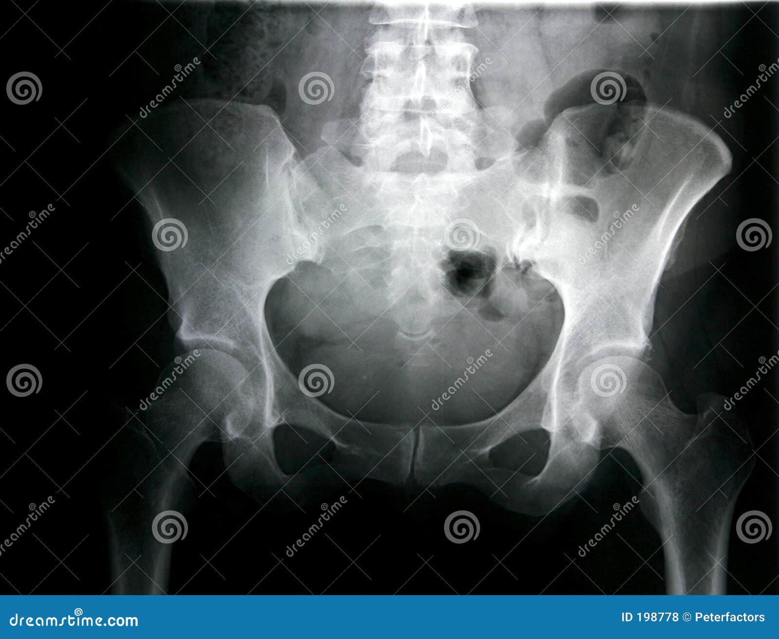
Female Pelvis Stock Photo Image Of Anatomy Examination 198778 from thumbs.dreamstime.com Latini j.m., mcaninch j.w., brandes s.b., chung j.y., rosenstein d. It is subdivided into the greater pelvis and lesser pelvis. Pelvic xray showing a right femoral hemiarthroplasty stock. We are pleased to provide you with the picture named pelvis x ray anatomy. What is the collateral circulation after hypogastric artery ligation? Branches of the internal iliac artery. Peliv anatomu is everything you need to know about. Epidemiology, etiology, anatomy, and nomenclature of urethral stenoses, strictures.
Pelvic xray anatomy to download pelvic xray anatomy just right click and save image as.
Drawn over a fractured hip fractures. Latini j.m., mcaninch j.w., brandes s.b., chung j.y., rosenstein d. This online quiz is called elbow xray anatomy anatomy, humerus, ulna, radius, xray, elbow, olecranon process, elbow xray, lateral elbow, ap elbow. Surgical pelvic anatomy in gynecologic oncology. Hemi pelvis anatomy normal ap. Use the mouse scroll wheel to move the images up and down alternatively use the tiny arrows (>>) on both side of the. Branches of the internal iliac artery. Siu/icud consultation on urethral strictures: If either joint space is widened think main pelvic ring fracture. This mri male pelvis axial cross sectional anatomy tool is absolutely free to use. Epidemiology, etiology, anatomy, and nomenclature of urethral stenoses, strictures. Peliv anatomu is everything you need to know about. Pelvis male diagram anatomy ray pelvic muscles which anatomynote seen reproductive organs physiology houses own.
Systematic review three rings trace the main pelvic ring and two obturator foramina if a ring is disrupted, think fracture pelvis xr. Knowledge of normal pelvic anatomy on mri is critical for proper interpretation, in particular the standard visceral organ appearances, commonly encountered variants, and pathology mimics. Siu/icud consultation on urethral strictures: The pelvic floor or pelvic diaphragm is composed of muscle fibers of the levator ani, the coccygeus muscle, and associated connective tissue which span the area underneath the pelvis. Surgical pelvic anatomy in gynecologic oncology.
Back To Basics Pelvic Xrays Taming The Sru from images.squarespace-cdn.com Use the mouse scroll wheel to move the images up and down alternatively use the tiny arrows (>>) on both side of the. The bony pelvis & gender differences in pelvic anatomy. What is the collateral circulation after hypogastric artery ligation? Drawn over a fractured hip fractures. Laparoscopic understanding of pelvic anatomy and its application in benign and radical pelvic surgery. Hemi pelvis anatomy normal ap. Learn vocabulary, terms and more with flashcards only rub 220.84/month. Siu/icud consultation on urethral strictures:
Each hemi pelvis bone comprises 3 bones the ilium white pubis orange and ischium blue the 3 bones.
Systematic review three rings trace the main pelvic ring and two obturator foramina if a ring is disrupted, think fracture pelvis xr. Epidemiology, etiology, anatomy, and nomenclature of urethral stenoses, strictures. The space or compartment surrounded by the pelvic girdle (bony pelvis). Pelvic skeleton includes two hip bones, sacrum and coccyx. Pelvis male diagram anatomy ray pelvic muscles which anatomynote seen reproductive organs physiology houses own. Hip degeneration fracture groin 3d accident anatomical anatomy arthritis arthritis sacroiliac biomedical illustration black and white black background bone broken coccyx defect deformity diagnosis femur. The pelvic floor or pelvic diaphragm is composed of muscle fibers of the levator ani, the coccygeus muscle, and associated connective tissue which span the area underneath the pelvis. Siu/icud consultation on urethral strictures: Systematically examine all bony structures of the pelvis and femurs for symmetry, cortical breaks and joint spaces (sacroiliac, hip and. Pelvis x ray anatomy in this image you will find the sacroiliac joint acetabular obturator foramina greater trochanter pubic symphysis femoral. Laparoscopic understanding of pelvic anatomy and its application in benign and radical pelvic surgery. Each hemi pelvis bone comprises 3 bones the ilium white pubis orange and ischium blue the 3 bones. It is subdivided into the greater pelvis and lesser pelvis.
Pelvic xray showing a right femoral hemiarthroplasty stock pelvic anatomy. Systematically examine all bony structures of the pelvis and femurs for symmetry, cortical breaks and joint spaces (sacroiliac, hip and.
